Tuesday 6 December 2011

Neuropsychological Assessment ( Week 11)

The last lecture of term talked about neuropsychological assessments help to identify and understand the exact brain damage on a intelligence scale and how much motor, emotional and other life treating problems are occurring at the same time as well as  accurate interpretation of the patients deficits and how difficulties these impairments are. There are different classes of neurolopsychological test, such as testing a patients intelligence, his learning and memory and others like testing a patients decision making. Many previous studies with these test have been done and one that is particularly interesting for me by Owen et al. (1997) examined  patients with Parkinson's disease that were either under the influence of medication or not, whether their spatial,verbal and visual woking memory was impaired. It was found that in all three tasks, patients that were medicated for Parkinson's disease did very badly,whereas patients with fewer symptoms did only poorly on the spatial working memory task. Compared to the non-medical patients who were fine on all three tasks. Previous studies suggested that especially cognitive deficits such as memory develops and progress within Parkinson's disease to the frontal lobes. This research agrees with previous material, that due the development of dopamine in the striatum the level of spatial working memory can be impaired.

One of the famous recognition memory task is the "Rey- Complex Figure" (Rey, 1941) which examines visual memory in patients with brain damage. Patients are asked to copy the figure first and that after a pause draw the figure from their memory. The reason why patients have problems with such task is that not just the memory is needed, also planning and attention is needed. The implicit memory of the patient is impaired as immediate and delayed conditions are used for this task. The reason for the non color use is that patients would be more able to recognize on the color rather than the figure. 

Also know as the "Complex Figure test", this shows a
clear impairment of the patients visual memory, by trying
to remember the figure

Tuesday 29 November 2011

Memory (Week 10)

While speaking last week about the frontal lobes and their involvement in memory task, this weeks lecture covert Memory in general. Memory is a very individual process that gives us the ability to retrieve, process and store information from our past and plan for the future. Different parts of memory are responsible in the way we make decisions, judgement and know things we can recall from the past. The primary memory, today know as short-term memory and the secondary memory, known as long-term memory, are found by James (1890). Our primary memory is only able to store temporally learned information, things we randomly pick up, whereas the secondary memory can be seen as a storage for varies things, it is assessed with our conscious, while the short-term memory is picked up due to our unconscious. 

Research done by Teng and Squire (1999) examined an amnesic patient who lost the ability to memorize  due to bilateral damage on his hippocampus but was able to remember back his lost places after looking at the spatial layout of places he used to go to. That proves that hippocampus that the medial temporal lobe are necessary for declarative memory which is long-term memory. Another very important aspect of the long-term memory, and a field where much research has been done, is the difference between the non-declarative part of memory, this procedural memory is meant by skills you have learned a long time ago and can recall them whenever you like, such as swimming or riding a bike. Memory one of the major systems in the human body that makes us unique in many ways and differs us from others. 


The Human memory is like a mind map that can be accessed at any point in time, we also use that type of structure to identify problems, work out things that are important, as well as make our "mind up" about something. 

Friday 25 November 2011

The executive functions of the frontal lobes


This weeks lecture covert the frontal lobes and its executive functions. The human species has the most developed frontal lobes, and it is also the last area to develop in children. The frontal lobes are involved in thinking and the connection between sensory and memories are crucial to achieve cognition. Problem solving and judgment are also undertaken by the frontal lobes that besides that have the capacity to plan ahead. Numerous tests have been done to measure frontal lobes functions, which in general assess the ability of the patient to arrange previous experience reasonably, rationalize and be able to behave impulsively. 

The executive functions of the frontal lobes embrace components such sustained attention where the individual cant deal with more than one things at a time, set- shifting and others. Set- shifting was especially measures in the Wisconsin Card Task (Milner, 1964) that demands a surprising change of rules (color or number). This means that patients with prefrontal cortex damage are failing to complete sorting the cards correctly. For patients with lesions, life is dependant on others, planning ahead is impossible and fundamental task like decision-making are affecting their behavior and personality and loosing track of organization is getting more complicated. 

The Wisconsin Card Task, where the patient has to ignorer the color and tell what it is. Sometimes you have to answer whatcolour the card has, when the scientist changes the rules. depending on the patients impairment, there are more or less problems in shifting from one task to another. 


Scientists like Banich (2009) are trying to relate neurobiological and psychological methods further by looking at brain tasks individually and differentiate the diverse neurological models to understand the executive functions from a more rational perspective. Executive functions are also crucial for intelligent behavior, are necessary for the theory of mind as the frontal lobes are involved in everything to be aware and conscious. 

Thursday 17 November 2011

Hemispheric lateralisation (Week 7)

This weeks lecture was about the hemispheric lateralisation of the human brain. The human brain is divided in two parts, the right and the left hemisphere, connected by the corpus callosum. Both hemisphere structures are identical to each other, however their functions are managed in different terms. It can be identified that the right hemisphere is dealing with judgement and decision making, such as including the age,spatial skills, facial properties and is also involved in contextual language and numerical computation (e.g. estimation and number comparison). Whereas the left hemisphere has been identified as a less inferior part on vocal tasks, on how our speech develops, the processing of speech and grammar/vocabulary. Today knows as Broca's area, that was found in 1861 by Pierre Paul Broca who distinguished after his patients death that because of syphilitic lesions in his left hemisphere the speech production of his patient could never develop. Later theories about the asymmetry of the brain has brought attention towards the brain lateralization that explains why most people are right handed and less are left handed. Studies on split-brain patients have been done which shows that when looking at something, the right hemisphere is active and the information, travels to the left side to pursue language and patients correctly identified what was on the screen. Now when information was presented to the left side of the hemisphere, the information travels to the right side of the brain, which does notch control language, and that proved that patients could not identify the object presented correctly.  So it seems like that split-brain patients have two brains, which is for us one brain with both sides different functions, seems for those patients more like two different systems not working together, and can deal with task separately and in different ways.   


This picture just shows the brain split into two parts and makes clear how the left hand side is responsible for information given on the right and how the right hemisphere deals with processing on the left. 

During the seminar this week we learned about the experiment that was based on Levy, Heller, Banich, & Burton (1983) first experiment, which is now replicated by  Rueckert (2005). I did the experiment myself and found that when being presented with the two chimeric faces, i had to press a key for which i thought was younger or older. Without even knowing which one to pick, unconsciously i pressed one key. The truth was that the two chimeric faces were always the identical mirror images of each other. So I gained a 100 % certainty when picking the image, my left hemisphere was working and i am right handed.  

Friday 11 November 2011

Emotion (Week 6)

Emotion, a quit simple term but hard to define. In this weeks lecture and seminar we discussed the different characerizations of emotions and how humans get emotional. Many previous done research suggests that the amygdala is responsible for our emotions (Adolphs, 1999). The amygdala is a small part of the hippocampus that takes in information that is provided by our senses and experiences from the outside world and  processes these information. The amygdala is linked to the higher cognitive processor  and the lower cognitive processor, also know as hypothalamic and brainstem systems. It can be said that based on all the cognitive functions the amygdala has been given, a emotional reaction is the outcome. However it can still be argued that the amygdala is not the only important system that is related to the retrieving and processing of information for our emotions. The additional reading i did for this week, Adolphs (1999) also suggested that the amygdala is responsible for decision-making and the dealing with memory. 

Information retrieved from: 
Adolphs, R. (1999). The Human Amygdala and Emotion. Neuroscientist, 5: 125

A very good video that helped me to understand the different theories on how our emotion is retrieved and processed in the brain:


In the seminar, we did a fantastic experiment on emotion. Viewing some pictures of faces, that showed an emotion and afterwards stating what emotion that was. It was really interesting to to such experiment, which i had never done before. afterwards we discussed the gambling task, which we had to perform before too. The gambling task, and so was about that there were four deck cards and you were asked to make the most money by choosing in each round one card, however some of the cards also carry a negative outcome, like 50 US Dollars minus the money you had already gained. after a while I did notice though that if you either press A or C you gain money and if you press B or D you would loose, yo automatically, because i want to win, kept on pressing A, which made me very happy. The outcome of this experiment, which had been replicated by a few researches showed that decision-making is an important process of emotions, either conscious or unconscious. It can be argued if emotions are necessary to make such decision or decisions in general or if it would be better to see everything from the outside and have no emotional response to things at all.

Wednesday 26 October 2011

Movement Disorders (Week 5)


The human movement is the basic framwork of the prefrontal and premotor cortex. Different routes in our brain are responsible for diverse actions that create control of thought and cognition. Movement is controlled by our spatial reference frame that involves knowledge about the location of the body in space and of course about the object you are paying attention for, such as grasping for a pen, your eyes focus on the object, your hand reaches out and makes the correct hand movement to grasp for it, this process is also called as sensory-motor transformation. Our premotor areas, are mainly dealing with internal and external events from our day to day life, also involved in specific goal targets, also called achievement, these are lying to the back of the prefrontal cortex. The prefrontal cortex is occupied with tasks like planning and decision-making, paying attention and deals more with general actions than object specific. The occipital lobes is involved with our visual system, where projections from the retrina are received where different types of neurons provide certain information for colour, orientation and motion. Two important pathways are responsible for our conscious information, the dorsal stream transmits to the to the parietal lobes and identifies where objects are located and the ventral stream provides information to the temporal lobes and categorizes what objects are. If damage to the ventral stream has occurred, visual agnosia can develop and also impairment in identifying objects, colour etc is highly likely. Another form of damage are movement disorders, that occur due to damage in the left hemisphere in the brain and cause physical impairment of movement, like gestures and grasping. If there has been damage to the Basal Ganglia, impairments in movement are expected. Different disorders are Parkinson’s, Huntington's and apraxia. Research has found that most patients with practical and dissociative impairment deal with more than one symptom, and it can also be said that that when a patient deals with more indications of the illness it might be quit mysterious how these come together, such as walking problems, twitches and uncontrollable arm movement called tremor. 


This model of movement disorders I'll find enormously helpful, to identify, of what brain area the disorder comes from  and also the yellow and red markings help to understand which ones are "positive" and "negative. Whereas positive and negative are the wrong terms to use, as its too extreme, all disorders are negative, however some have further damage than others.  

For the movement disorder, Parkinsons Disease what mainly affects elderly people, I have found a really good and interesting article by BBC, which made me smile today. I felt that even within all these sad disorder theories, a surgery of an elderly man with parkinsons disease was successful and he has 95% of his life back:


I also have found an interesting page towards functional movement disorders, that support people with the disorder and help to adapt to the real world, as psychological seen some patients believe that the disorder develops as they would have done something wrong in their life, which is not true. 


    http://www.neurosymptoms.org/#/movement-disorders/4533053142


I have read the paper about, "Abnormalities in the awareness of action" by Blakemore,Wolpert and Frith (2002). A particular model was designed to establish motor learning and control in individuals to identify which parts of our motor control are responsible for the human awareness. It has been found that individuals not always have conscious motor control of things. Such as picking up a pen, you are targeted towards the pen, and already make the correct hand movement, grasping for it. So you are consciously aware for that but unconsciously there goes far more work into one movement, like strengthen and loosing your muscle tone. Abnormalities in movement are described as a dismissed control of action. One of the abnormalities is called "optic Ataxia", I´ll find that one a slightly confusing as i thought the patient would not be able to see the object properly, but actually the subject has problems grasping the item, so the individual can see quit good, just cannot focus completely on the object. However that does not mean if you character wise a bit clumsy that you have a disorder. Another abnormality, I´ll find quit fascinating is the "utilization behavior" which means that patients use some objects at the wrong time, like it is raining outside and the patient wants to wear its swimming costume, which obviously is not really wrong but in that moment, it would be used inappropriately. However there are some other abnormalities that are being discussed in the paper and i have also read some more about them, I do feel that the model itself and topics addressed by the researcher are sometimes controversial, as not enough evidence has been found to what circumstances does our awareness becomes more central for the model, to conclude the paper, I feel that some more research has to be done and belief that neurological and psychiatric factors have to be more systematically evaluated to complete the model to understand the main principles of motor control and awareness. 

Wednesday 19 October 2011

VISUAL PERCEPTION (DISORDER PART 2)

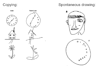
Spatial neglect also talked about as unilateral spatial neglect or hemi-inattention, that is a seizure to failing respond to events in the hemifield contralateral to a previous happened damage to the brain. The more common one of neglect is the visual neglect, which occurs to most patients after having a stroke. Patients tend to see only half of the world, depending on where in the brain the lesion is, e.g. the patient does not acknowledge the left side of the world, as he has right hemisphere damage in his brain. 


This is a classic example of a "copying" and
"spontaneous drawing" task by a patient with visual neglect
of the brain. In the copying task, it shows clearly, the denial
of the left world, and even in the spontanious drawing
task, the patient ignores the fact that there is a left piece to
his drawings.  
Patients that have spatial neglect “ignore” the other half of the world in a way that many experiments on measuring eye movements, line bisection, where the patient is asked to place a mark  in the centre of a straight line, here it is assumed that the patient with neglect, moves either more to the right, or when the lines are vertical, the patients drawings tend to move away from the body. Another experiment that has been done was, where the patient was asked to find all the Ts within a letter mix of Ts and Ls. The patient with the neglect on the right side does not know that the left side even exists anymore. Interesting is also the anatomy of the neglect as I believe that the parietal lobe not always played a critical role in human life. Research suggests that the parietal lobe actually nothing to do with the spatial neglect, researcher are more concerned with the temporal lobe and also focal lesions of the right inferior frontal lobe can lead to spatial neglect, therefore it can be said that the biological theories on where neglect actually takes place are controversial. Tests have been done on monkey’s brain that revealed that direct attention and eye movements to a three-dimensional target are linked with neuronal responses due to their dorsal visual stream, which is part of the parietal cortex that can be critical towards spatial action and vision, the same is expected in human individuals where lesions of the parietal cortex can be related with spatial deficits. Neglect is modulated. There is complexity of the visual scene and patients with lesions in their right hemisphere also have weakened recognition on their right side. 


In terms of the deepness of the hemineglect, there also dysfunction of memory in patients, where the patients where asked to look at a map from the cathedral in milan and describe what they see when looked at the front of the church, patients only described the right part of the map, then when the examiner turned the map around and asked the patient to describe what they see by looking at the cathedral from the back, they also described the right part, which i think is quit funny in terms of that, that is the actual side their brain " abandoned" before and never acknowledges that, that side even exists. also what i just realised, when a patient has a neglect for the left side, what happens when the patient would turn its head, wouldn't he than see what he actually missed the whole time, or is it like a circle that even though he does turn, his view on the world and what exists and what doesn't exists stays the same?